Polymers in Medicine

Polim. Med.
Scopus CiteScore: 3.5
MNiSW - 70

ISSN 0370-0747 (print)
ISSN 2451-2699 (online) 
Periodicity – biannual

Download original text (EN)

Polymers in Medicine

2026, vol. 56, nr 1, January-June, p. 7–17

doi: 10.17219/pim/218616

Publication type: review

Language: English

License: Creative Commons Attribution 3.0 Unported (CC BY 3.0)

Download citation:

  • BIBTEX (JabRef, Mendeley)
  • RIS (Papers, Reference Manager, RefWorks, Zotero)

Cite as:


Tahir M, Pettineo S, Vicini S, et al. Sodium alginate for wound healing applications: A review. Polim Med. 2026;56(1):7–17. doi:10.17219/pim/218616

Sodium alginate for wound healing applications: A review

Muhammad Tahir1,A,B,C,D, Simone Pettineo2,A,B, Silvia Vicini2,A,E,F, Maila Castellano2,E,F, Marina Alloisio2,E,F, Ketul Popat3,E,F, Harishkumar Madhyastha4,E,F, Kaoru Ohe5,E,F, Kentaro Sakai6,E,F, Alina Sionkowska1,A,E,F

1 Laboratory for Biomaterials and Cosmetics, Faculty of Chemistry, Nicolaus Copernicus University in Torun, Poland

2 Department of Chemistry and Industrial Chemistry, University of Genova, Italy

3 Department of Bioengineering, George Mason University, Fairfax, USA

4 Department of Cardiovascular Physiology, Faculty of Medicine, University of Miyazaki, Japan

5 Center for Science and Engineering Education, Faculty of Engineering, University of Miyazaki, Japan

6 Division for Research Facility Support Organization for Promotion of Research and Industry – Academic Regional Collaboration, University of Miyazaki, Japan

Graphical abstract


Graphical abstracts

Abstract

Sodium alginate (SA) is a polysaccharide biopolymer widely used in wound healing applications due to its beneficial role in the hemostatic, inflammatory, and proliferative phases of tissue repair. Its hydrophilic nature supports the wound healing process by enabling efficient absorption of wound exudate and maintaining a moist microenvironment conducive to tissue regeneration. Moreover, SA can form highly porous structures that promote oxygen diffusion and provide a suitable scaffold for neovascularization and new tissue formation. This review summarizes recent advances in the application of SA in wound dressings. A bibliometric analysis of Scopus data using the keywords “sodium alginate” AND “wound healing” reveals a growing number of publications in recent years, highlighting the increasing scientific interest in this field. The expanding utilization of SA in wound healing may be attributed to its favorable properties, including biocompatibility, biodegradability, and low toxicity.

Key words: wound healing, sodium alginate, biopolymer, alginic acid, porous materials

Introduction

Biopolymers can be extracted from bio-based materials using various techniques. They can also be produced biotechnologically or synthesized directly from building blocks derived from living organisms. Thus, in general, nature is the primary source of biopolymers.1 Jabeen and Atif defined biopolymers as macromolecules that are primarily composed of monomeric units of natural origin.2 Biopolymers are advantageous due to their environmentally friendly nature.3 Nitta and Numata discussed the classification of biopolymers, which are mainly divided into 3 types: polysaccharides, nucleic acids, and proteins.4 In the huge family of polysaccharides, one can find starch, cellulose, chitin, chitosan, and many more, for example, alginate.5

The primary sources of alginate reported are brown seaweed and marine algae.6, 7 Alginate has a unique non-toxic nature and biocompatibility, which make this biopolymer attractive for biomedical and cosmetic applications.8, 9 It exhibits bioadhesivity, which is the primary reason for its widespread use in pharmaceutical applications.10 It may form sponge-like structures that are capable of absorbing water.11 Alginate-based hydrogels are formed through ionic crosslinking.12 Salarvand et al. reported the presence of hydroxyl and carboxyl groups in alginate, which gives the possibility of hydrogen bond formation.13 Moreover, alginate contains negative ions capable of further interactions.14 Wang et al. reported that sodium alginate (SA) and its oxidized derivatives show good cytocompatibility.15 Alginate-based nanofibers are widely utilized in biomedical applications, especially in tissue engineering and wound healing.16,17 It has recently been observed that SA applications in various areas are growing, particularly in wound healing.

Sodium alginate

The properties of SA are widely studied. Phùng et al. mentioned the water solubility of SA,18 whereas Yan et al. reported that SA is insoluble in ethanol.19 Sodium alginate is highly preferred in several applications due to its outstanding ability to form films.20,21 Yan et al. described SA as a low-cost polymer presenting high potential.19 The presence of functional groups, such as hydroxyl and carboxyl groups, makes it easy to modify.22 Belalia and Djelali reported the rheological behavior of SA. In their research, it was concluded that it presents shear-thinning non-Newtonian flow behavior in its aqueous form.23 Viscosity was reduced with an increase in shear rate; this behavior has been reported as shear-thinning behavior in the literature.24

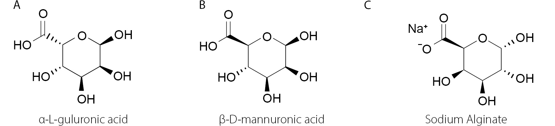
In the structural formula of SA, the main constituents are α-L-guluronic acid and β-D-mannuronic acid.25,26 ChemDraw 25.0.2 software (Revvity Signals Software) was used to draw the structural formulas of SA and its constituents, which are presented in Figure 1. The “Structure” tab of the ChemDraw software was selected, and in this tab, “Convert Name to Structure” was chosen to draw the structural formulas of SA and its constituents. The names used are also presented in the figure alongside their corresponding structural formulas. Therefore, in short, SA is a combination of 2 uronic acids. Sodium alginate sourced from algae contains a significant portion of other cations: Ca (calcium), Mg (magnesium), and Sr (strontium), which are treated with hydrochloric acid (HCl) to obtain alginic acid through a liquid wash mechanism.27

The addition of sodium carbonate (Na₂CO₃) leads to a pH range of 9–10. Na₂CO₃ can also be replaced with alternatives such as sodium hydroxide (NaOH), sodium bicarbonate (NaHCO₃), or sodium chloride (NaCl). This leads to the conversion of alginic acid (which is water-insoluble) to SA (a water-soluble form).28 It can be followed by either the Ca-alginate or the alginic acid route. The route considering alginic acid involves the treatment of SA with HCl. The 2nd route is the Ca-alginate route, which involves the addition of calcium chloride to SA to form Ca-alginate.29 Ca-alginate is further treated with HCl to obtain alginic acid. The alginic acid obtained from these 2 routes is further treated with Na₂CO₃ to produce SA in its pure form.30

Porosity of sodium alginate materials

Sodium alginate materials can be fabricated into gels. Abka-Khajouei et al. reported that alginate gels presented pores, with the pore size specified in their research ranging from 5 nm to 200 nm. They mentioned electron microscopy for pore size demonstration.31 Da Silva et al. reported that as the SA concentration increases, it causes a decrease in the material’s porosity. Therefore, an inverse relationship between SA concentration and material porosity was presented. Porosity in wound dressings is preferred because it facilitates the process of oxygenation.32 Sinha et al. reported that wound dressings with the highest porosity had the greatest tendency to absorb wound exudate.33 Liu et al.34 also demonstrated that SA gel presented pores. They conducted field emission electron microscopy (FESEM) on SA gels with the addition of calcium ions (Ca2+) to demonstrate the pores.

MATLAB and Python are referred to as programming languages.35,36 They are effectively utilized for the visualization and demonstration of pores. Jenkins et al. were the developers of PoreScript (a MATLAB algorithm), and PoreScript was used to determine pore size from scanning electron microscopy (SEM) images.37 Polez et al. utilized SEM images to determine porosity using PoreSpy,38 a Python package that characterizes porous materials from 3D images.39 Negut and Bita reported the use of a convolutional neural network (CNN) to identify pore size distribution in hydrogels.40 Zhang et al. reported CNN as a deep learning model.41 El Naqa and Murphy described deep learning as a sub-classification of machine learning, and stated that deep learning has the capacity to be trained from raw data.42 Machine learning has been referred to as a field within artificial intelligence.43 Three types of machine learning are reported in the literature: supervised learning, unsupervised learning, and reinforcement learning.44,45

Supervised machine learning utilizes labeled data.46 Unsupervised learning is based on unlabeled data.47 Reinforcement learning allows an agent to take actions.48 Correct actions in reinforcement learning lead to earning rewards, whereas wrong decisions result in failure.49 Saberian et al. utilized ImageJ software (National Institutes of Health (NIH), Bethesda, USA) to measure pores based on SEM images. The hydrogel utilized in their research was based on alginate/chitosan and also included honey and aloe vera. The prepared hydrogel was intended for use as a wound dressing.50 Olevsky et al. developed PoreVision using a Python script. PoreVision utilizes Python libraries, primarily OpenCV, NumPy, and Pandas. It was used to measure pore size from SEM images and can be utilized for gels and porous materials, as indicated in their research.51 Shkarin et al. developed quanfima, a Python-supported package that can be used to analyze the morphology of biomaterials, including porosity.52 MICPY has been highlighted for pore analysis and has been reported as a Python-supported package. It can analyze SEM images.53,54 Nair et al. utilized CTAnalyzer software for pore size determination, which was made possible by using a 3D object analyzer within the software.55 Karaca and Aldemir Dikici utilized a deep learning model, which they named Pore D2. In their study, YOLOv5 was used for object detection, and SEM images were utilized as the input. EasyOCR has been used to extract text from images, and it is a Python library that employs a deep learning approach. The output layer provided accurate information on pore size.56

Wettability of sodium alginate materials

The hydrophilicity of the material’s surface may be a reason for better cell adhesion. Yuan and Lee presented the relationship between the water contact angle and wettability in the context of liquid–solid interaction. According to their research, contact angles below 90° exhibited the highest wettability. A contact angle greater than 90° was a clear indication of low wettability.57 Kumar and Prabhu described reactive and non-reactive wetting. A reaction occurring between the spreading liquid and the substrate material impacts the mechanism of wetting, which is reported as reactive wetting. If no reaction occurs between the spreading liquid and the substrate material, it is reported as inert or non-reactive wetting.58 Xie et al. conducted contact angle analysis for membranes made of SA, and SA 100 and SA 200 were named for the lower amounts of guluronic acid. Manugel DMB was named for a higher amount of guluronic acid. A Milli-Q water drop was utilized to obtain contact angle measurements, and the analysis was conducted on alginate–chitosan–alginate (ACA) membranes. The water contact angle for SA 100, as reported in their study, was 65.9 ±1.0°. SA 200 presented a water contact angle of 68.5 ±2.4°. The water contact angle of Manugel DMB recorded in their research was 77.7 ±3.3°. These values are reported as mean ± standard error (SE). The number of analyses specified in their study was 3. In short, their results showed that an increase in guluronic acid content led to a reduction in hydrophilicity.59 Karmakar et al. analyzed the contact angle of a hydrogel composed of SA and carboxymethyl cellulose in a 1:1 ratio, reporting a contact angle value of 48.32 ±1.5°. Contact angle measurements were recorded using distilled water, and 3 measurements were taken to obtain the mean value.60 Mujawar et al. conducted contact angle measurements on 3D-printed samples, including those made from SA–gelatin and SA–gelatin–Aloe barbadensis extract. Six contact angle measurements were taken using water as the probe liquid. The recorded water contact angle for 3D-printed SA–gelatin was 42.31 ±3.23°. The water contact angle reported for SA–gelatin–Aloe barbadensis extract was about 53.2 ±4.7°.61 Sisakht et al. prepared a hydrogel with a 50:50 weight ratio of SA and poly(acrylic acid). They utilized both SA and poly(acrylic acid) at a concentration of 2% wt/vol. They also analyzed the contact angle in combination with fibroblast growth factor (FGF1). Deionized water was used as the probe liquid for the contact angle analysis. Three measurements were recorded for the samples. The contact angle reported for SA was 57.87°. In the case of the SA–poly(acrylic acid) hydrogel, the reported contact angle was 18.52°. The SA–poly(acrylic acid) hydrogel in combination with FGF1 exhibited a water contact angle of approx. 62.56°.62 Hosseini et al. prepared a hydrogel composed of carboxymethyl cellulose and SA. Subsequently, they combined it with simvastatin, resulting in a final form known as the carboxymethyl cellulose/SA–simvastatin hydrogel. The recorded water contact angles for carboxymethyl cellulose–SA and carboxymethyl cellulose/SA–simvastatin were 43.3 ±2° and 59.1 ±4°, respectively. They utilized water as the probe liquid, and 3 measurements were recorded for each sample.63 Table 1 presents a summary of the contact angle measurements for SA-based materials. Isa Rahim et al. specified that contact angles lower than 90° were indicative of hydrophilicity, and contact angles between 90° and 150° were indicative of hydrophobicity.64 Chen et al. specified that a surface with a contact angle higher than 150° can be reported as superhydrophobic.65 Choi et al. reported the use of SA for wound dressings, and the reason given in their study was its hydrophilicity.66 Jin et al. examined the impact of hydrophilic polymers on swelling, and their study concluded that SA presented the maximum swelling among the analyzed hydrophilic polymers.67 Feng and Wang highlighted the use of hydrogels with maximum swelling capacity for wound healing due to their ability to absorb wound exudates.68 Singh and Pramanik reported better cell adhesion for polymeric films composed of SA and chitosan than for alginate itself, and it was also mentioned in their study that the contact angle was lower than 90°.69

Sodium alginate wound healing

Zahid et al. indicated that the use of SA for wound healing applications is promising due to its liquid absorption.70 The stages of wound healing have been demonstrated in various studies and include hemostasis, inflammation, proliferation, and remodeling phases.7173 Hemostasis is the first stage of wound healing, and in this phase, blood clot formation takes place.74 Periayah et al. explained the blood coagulation mechanism in significant detail. Their study indicated the key role of platelets in thrombus formation.75 Zhang et al. demonstrated that platelets play a vital role in vascular repair.76 Huang et al. reported that SA can enhance platelet concentration.77 Zhang et al. recommended SA for blood coagulation applications.78 Chen et al. reported in their study the preparation of a hemostatic powder, which involves the presence of SA, CaCl₂, graphene oxide, and cerium nitrate. The concluding remarks in their study indicated that the hemostatic powder presented the ability to absorb water, resulting in a shorter hemostatic time. The hemostatic powder effectively decreased blood loss and enhanced the wound healing mechanism due to the presence of Ce3+ (which acts as a free radical scavenger).79 Zhou et al. analyzed the blood coagulation of a gelatin sponge individually and in combination with SA, and their study summarized that blood coagulation was enhanced due to the presence of SA.80 Wang et al. prepared a powdered composite that included SA and poly(γ-glutamic acid) (PGA). The prepared powdered composite was found to arrest or block bleeding.81 Li et al. presented a material composed of SA, silk fibroin, and thrombin. It was mentioned that the prepared material could promote hemostasis.82 Xie et al. prepared a hydrogel containing SA, chitosan, and oxidized dextran. Their study results showed that the prepared hydrogel reduced blood loss, which significantly supports the basis for improved hemostasis.83 Khattak et al. mentioned that persistent inflammation was a significant reason for the slowing down or delay of the wound healing mechanism.84 Zhang et al. reported that inflammation was a notable challenge in wound healing.85 Liu et al. highlighted that the anti-inflammatory effect was essential for wound healing.86 Summa et al. prepared a wound dressing material composed of SA and povidone–iodine. The results of their study indicated that the prepared material exhibited an anti-inflammatory effect.87 Zhao et al. formulated a composite that included low-viscosity SA combined with titanium dioxide and temozolomide nanoparticles. Their study reported the anti--inflammatory effect of the designed composite. Their research summarized that the anti-inflammatory effect was enhanced due to the prominent presence of SA.88 Karmakar et al. initially prepared a SA and carboxymethyl cellulose composite, which was then crosslinked to a decellularized extracellular matrix. The crosslinker used in their study was calcium chloride, resulting in a final hydrogel composed of SA, carboxymethyl cellulose, and decellularized extracellular matrix. According to their research, the prepared hydrogel exhibited anti-inflammatory properties.60 Zhang et al. prepared a blend of SA and carboxymethyl chitosan, which was then loaded with curcumin. Sr2+ was used for the crosslinking of the polymers. The prepared hydrogel exhibited anti-inflammatory activity, leading to an enhanced wound healing mechanism.89 Raguvaran et al. prepared hydrogels composed of SA and gum acacia, which were incorporated with zinc oxide nanoparticles. Their research concluded that polymers in combination with zinc oxide nanoparticles tend to boost the proliferation of fibroblasts.90 Song et al. demonstrated that fibroblast proliferation is essential to promote the wound healing mechanism.91 Harper et al. presented fibroblast migration as a component of the proliferative phase of wound healing.92 Kibungu et al. reported that fibroblast cells assist in the production of collagen.93 AlShaali et al. also described that granulation tissue starts to form during the proliferation stage.94 Zhu et al. utilized a hydrogel based on SA, which was incorporated with Capparis spinosa L., and the incorporated hydrogel exhibited biocompatibility. It also boosted cell proliferation.95 Zhou et al. reported that enhanced cell proliferation encouraged wound healing.96 Ma et al. synthesized a nanocomposite based on SA, polyvinyl alcohol, and graphene oxide. Their research summarized that nanocomposites may result in enhanced cell proliferation.97 Dodero et al. prepared electrospun membranes of SA, which were crosslinked with divalent cations such as Ca2+, Sr2+, and Ba2+. In the final step, they prepared electrospun mats of SA loaded with zinc oxide nanoparticles, which were crosslinked with Sr2+ ions due to excellent cell adhesion. The electrospun mat prepared in their research was recommended for wound healing applications.98 Ding et al. utilized electrostatic spinning to synthesize a membrane based on SA and polyvinyl alcohol. Shikonin was incorporated into the electrospun membrane. It was found that the shikonin-loaded membrane boosted vascular endothelial growth factor A (VEGF-A) in the proliferative stage of wound healing.99 Liu et al. reported shikonin as a naphthoquinone pigment useful in the preparation of shikonin-based nanomedicine.100 Guo et al. highlighted Lithospermum erythrorhizon as the source of extraction for shikonin.101 Lin et al. referred to Lithospermum erythrorhizon as a Chinese medicinal herb.102 Tian et al. reported the antimicrobial and anti-inflammatory properties of shikonin in their research.103 Guo et al. mentioned the antioxidant and antithrombotic properties of shikonin.101 Ye et al. reported the anti-tumor effect of shikonin.104 Several studies reported shikonin as an ideal option for the treatment of diabetic wounds.105107 Eming et al. described VEGF-A in relation to angiogenesis in wound healing.108 DiPietro presented that angiogenesis results from hypoxia.109 In the case of injury, hypoxia is primarily responsible for the recruitment of hypoxia-inducible factors such as HIF-1. It supports the mechanism of angiogenesis during wound healing.110 Dawood et al. highlighted angiogenesis as a key feature linked to the proliferative phase of wound healing.111 Bahadoran et al. referred to the proliferative phase by another name, the growth phase of wound healing.112 Çerçi et al. utilized amoxicillin in combination with SA and polyvinyl alcohol. Polyvinyl alcohol was employed at a concentration of 12% wt/vol. Sodium alginate was utilized at a concentration of 1% wt/vol. The ratio selected for polyvinyl alcohol and SA was 2:1 (vol/vol) for the electrospinning solution, and 6.4 mg of amoxicillin trihydrate was added to the electrospinning solution. The electrospun nanofibrous mats were prepared by electrospinning. According to their research conclusions, the amoxicillin-loaded electrospun nanofibrous mat exhibited antibacterial properties. Based on their in vitro studies, the prepared nanofibrous mat loaded with amoxicillin was recommended for wound dressing applications.113 Kaur et al. reported amoxicillin as an antibiotic belonging to the penicillin class.114 Ayavoo et al. reported that the remodeling phase of wound healing was responsible for the conversion or transformation of fibroblasts into myofibroblasts.115 The remodeling phase of wound healing tends to remodel collagen III to collagen I.116 Yang et al. recommended a scaffold composed of SA, silk fibroin, and hyaluronic acid for extracellular
matrix remodeling.117

Bibliometric analysis

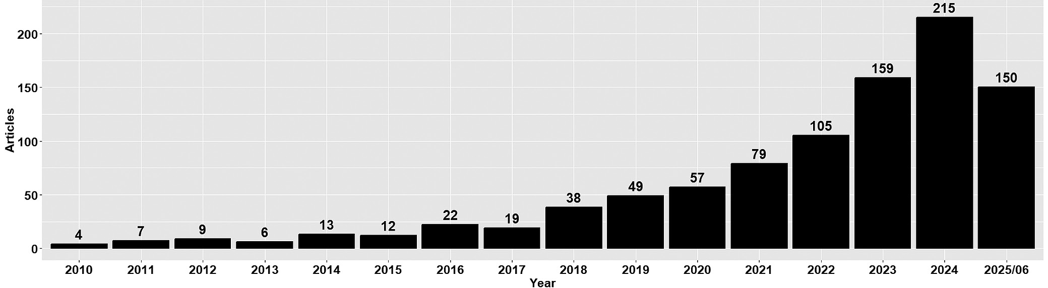
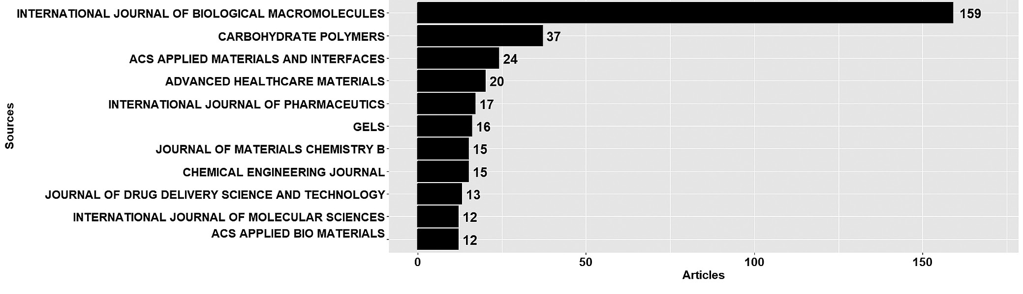
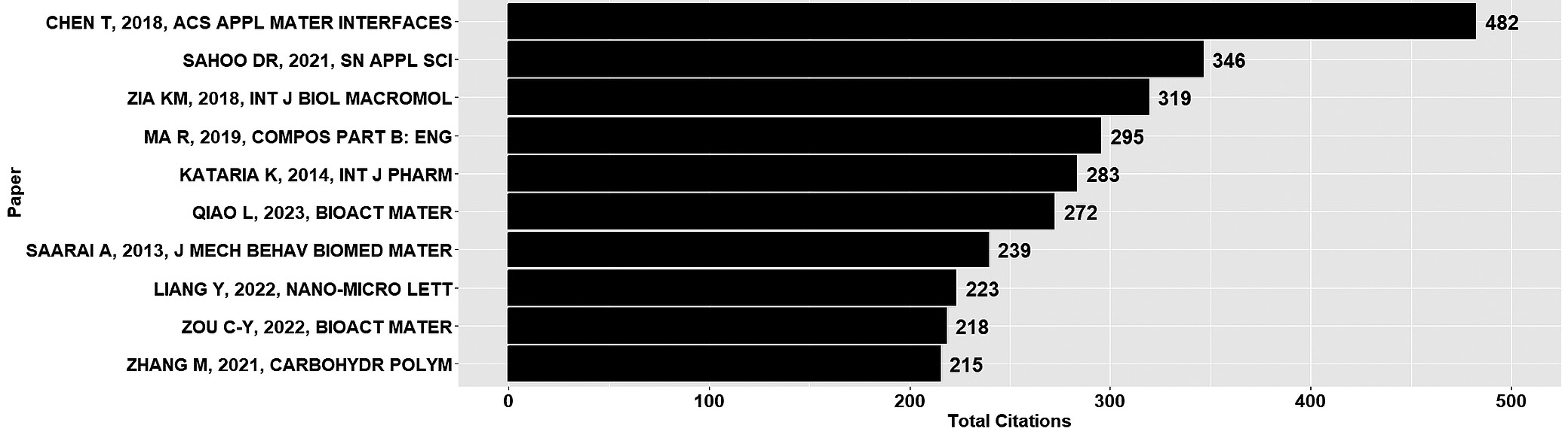
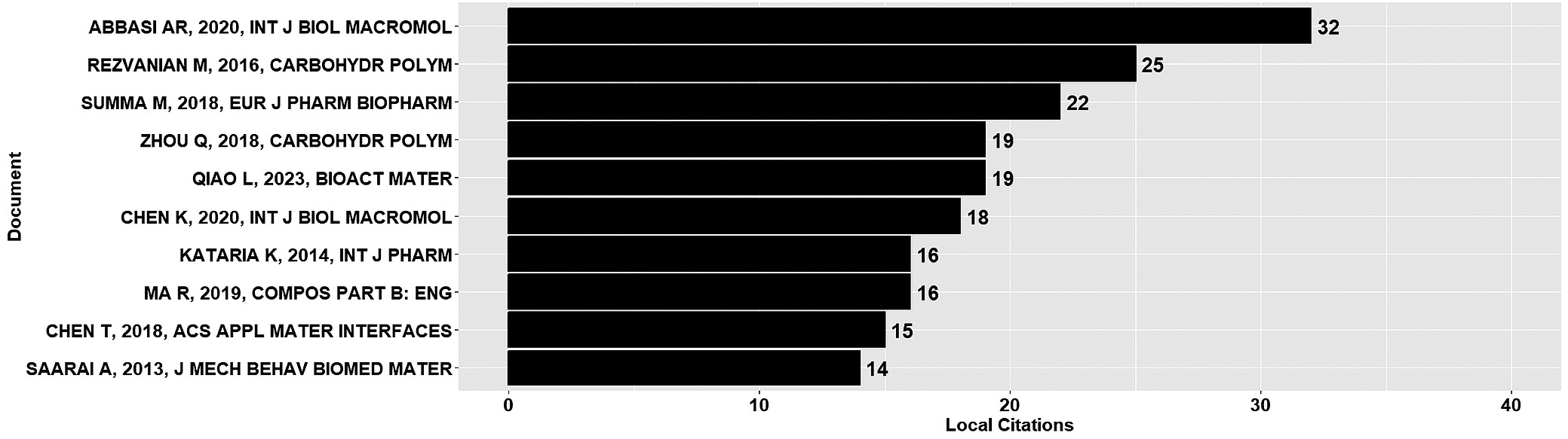
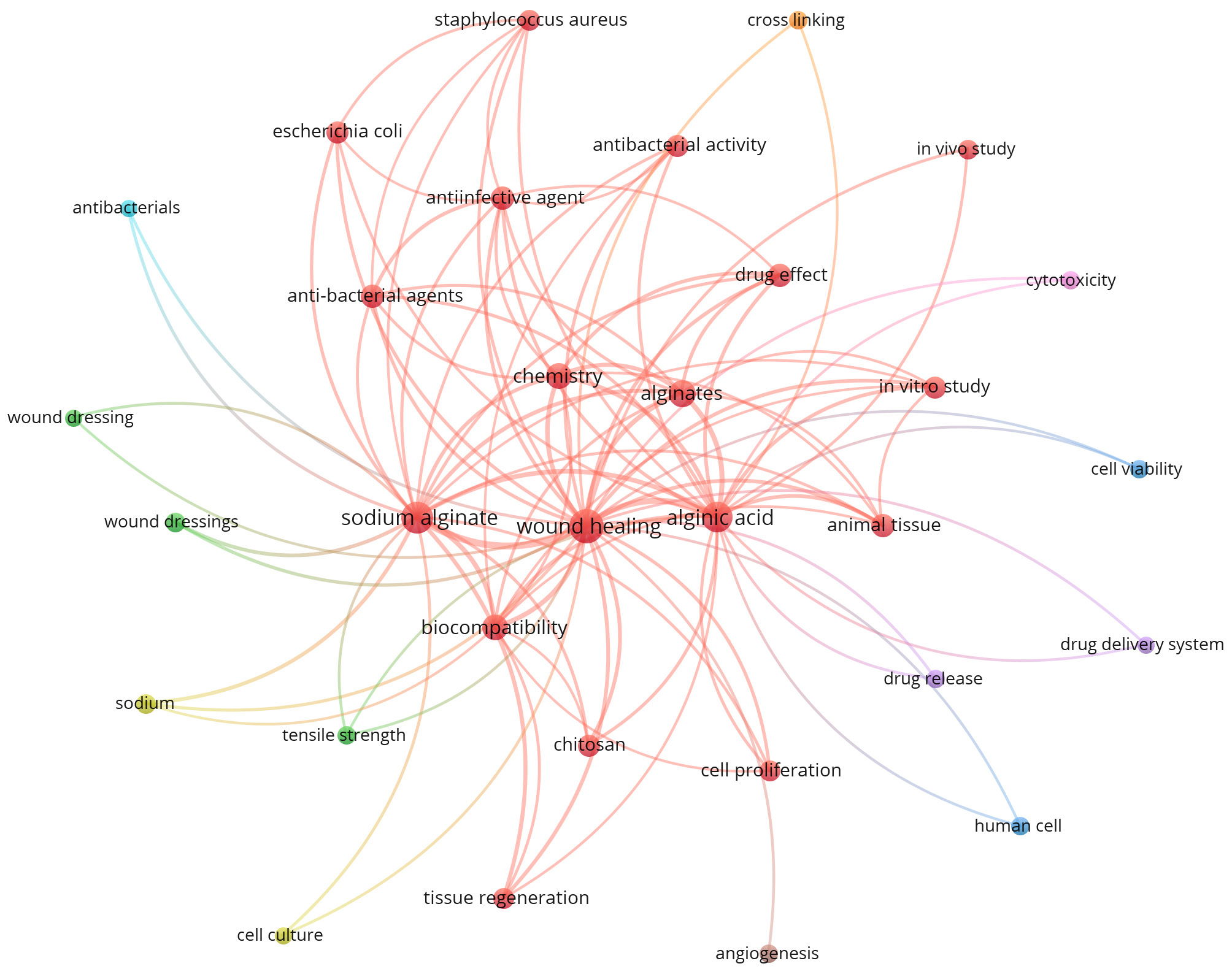
The bibliometric analysis was conducted using the R software v. 4.5.1 (R Foundation for Statistical Computing, Vienna, Austria) and VOSviewer (https://www.vosviewer.com/). Initially, the “bibliometrix” package was installed, followed by loading the library (bibliometrix). The biblioshiny() function was used to open the web-based interface for bibliometrix. Bibliometrix and biblioshiny were developed by Aria and Cuccurullo.118 The data were obtained from Scopus, and the document search was conducted using the terms “Sodium Alginate” AND “Wound Healing”, with the search restricted to “Article title, Abstract, Keywords” from 2010 to 2025 (access date: 27 June 2025). A total of 944 documents were generated from the search for “Sodium Alginate” AND “Wound Healing”. The annual scientific production data from Scopus for the specified search are presented in Figure 2. The analyzed data show that the number of articles from 2018 to 2024 increased continuously, indicating the growing prominence of the presented research in recent years. The most relevant sources are depicted in Figure 3. It represents the International Journal of Biological Macromolecules, the leading journal presenting pertinent articles, with 159 publications. The 2nd most relevant source was Carbohydrate Polymers, with 37 articles. The 3rd most relevant source was ACS Applied Materials and Interfaces, with 24 articles reported. The most globally cited papers were analyzed using Scopus data, and the results are presented in Figure 4, Figure which shows the papers with the most significant global citation impact. The impact of local citations was also investigated, and the highest number of locally cited documents was 32, as depicted in Figure 5. Van Eck and Waltman developed VOSviewer for bibliometric mapping.119 The Scopus data file was imported into VOSviewer, which helped identify relevant keywords through co-occurrence analysis. The co-occurrence analysis is presented in Figure 6.

Conclusions and future outlook

Several factors must be considered in the wound healing mechanism, including the hydrophilicity, porosity, and biocompatibility of wound dressing materials. Sodium alginate is primarily used in hemostatic powders for blood coagulation, as blood coagulation is the initial phase of wound healing. Wound healing is often delayed due to prolonged inflammation. Sodium alginate exhibits significant anti-inflammatory properties, facilitating early wound healing. The proliferation phase of wound healing is the combined result of angiogenesis, fibroblast migration, and collagen deposition. Sodium alginate can effectively promote the proliferative phase of wound healing by contributing to tissue granulation. Sodium alginate scaffolds also promote extracellular matrix remodeling. Machine learning is widely used to explore porous material structures. In the future, there is a need to develop an application programming interface (API) to facilitate data communication, particularly an API for the porosity of SA-based materials. Bibliometric analysis also revealed that open-access journals are focusing on promoting research related to SA wound healing applications. Although many papers regarding the application of SA have been published so far, it can be expected that more research articles on this material will be published in the future, as many research groups worldwide are working on alginate and its combinations with other
macromolecules.
Tables


Table 1. Contact angle measurements for sodium alginate (SA)-based materials

Sample

Number of measurements

Probe liquid

Contact angle

Reference

SA 100

3

Milli-Q water

65.9 ±1.0°

59

SA 200

3

Milli-Q water

68.5 ±2.4°

59

Manugel DMB

3

Milli-Q water

77.7 ±3.3°

59

SA–carboxymethyl cellulose (1:1 ratio)

3

distilled water

48.32 ±1.5°

60

SA–gelatin

6

water

42.31 ±3.23°

61

SA–gelatin–Aloe barbadensis extract

6

water

53.2 ±4.7°

61

SA (2% wt/vol)

3

deionized water

57.87°

62

SA–poly(acrylic acid) (50:50)

3

deionized water

18.52°

62

SA–poly(acrylic acid)–FGF1

3

deionized water

62.56°

62

Carboxymethyl cellulose–SA

3

water

43.3 ±2°

63

Carboxymethyl cellulose–SA–simvastatin

3

water

59.1 ±4°

63

Figures


Fig. 1. Structural formulas of (A) α-L-guluronic acid, (B) β-D-mannuronic acid, and (C) sodium alginate
Fig. 2. Annual scientific production (“Sodium Alginate” AND “Wound Healing” search within Scopus; access date: June 27, 2025)
Fig. 3. Most relevant sources with the number of articles (“Sodium Alginate” AND “Wound Healing” search within Scopus; access date: June 27, 2025)
Fig. 4. Most globally cited papers (“Sodium Alginate” AND “Wound Healing” search within Scopus; access date: June 27, 2025)
Fig. 5. Most locally cited documents (“Sodium Alginate” AND “Wound Healing” search within Scopus; access date: June 27, 2025)
Fig. 6. Co-occurrence of keywords (“Sodium Alginate” AND “Wound Healing” search within Scopus; access date: June 27, 2025)

References (119)

  1. Raina N, Rani R, Pahwa R, Gupta M. Biopolymers and treatment strategies for wound healing: An insight view. Int J Polym Mater Polym Biomater. 2022;71(5):359–375. doi:10.1080/00914037.2020.1838518
  2. Jabeen N, Atif M. Polysaccharides based biopolymers for biomedical applications: A review. Polym Adv Techs. 2024;35(1):e6203. doi:10.1002/pat.6203
  3. Thakur VK, Thakur MK, eds. Functional Biopolymers. Cham, Switzerland: Springer International Publishing; 2018. doi:10.1007/978-3-319-66417-0
  4. Nitta S, Numata K. Biopolymer-based nanoparticles for drug/gene delivery and tissue engineering. Int J Mol Sci. 2013;14(1):1629–1654. doi:10.3390/ijms14011629
  5. Ahmed HB, Mikhail MM, El-Shahat M, Emam HE. Clustering of carbon quantum dots from polysaccharides (cellulose, alginate, chitosan) versus heterocyclic compounds: Synthesis, characterization and medical applications. Carbohydr Polym Technol Appl. 2025;9:100738. doi:10.1016/j.carpta.2025.100738
  6. Shi C, Xu R, Hu W, et al. Fabrication of alginate dialdehyde-gelatin crosslinked hydrogels incorporated with cinnamaldehyde nanoparti-cles for meat preservation. Int J Biol Macromol. 2025;298:140063. doi:10.1016/j.ijbiomac.2025.140063
  7. Kulkarni VS, Butte KD, Rathod SS. Natural polymers–a comprehensive review. Int J Res Pharm Biomed Sci. 2012;3(4):1597-1613. Availa-ble from: https://www.researchgate.net/profile/Vishakha-gajrekulkarni/publication/236217541_Natural_Polymers-_A_comprehensive_Review/links/595b4913a6fdcc36b4daa3f7/Natural-Polymers-A-comprehensive-Review.pdf
  8. Al-Roujayee AS, Hilaj E, Deepak A, et al. Alginate-based systems: Advancements in drug delivery and wound healing. Int J Polym Mater Polym Biomater. 2025;74(9):846–874. doi:10.1080/00914037.2024.2375343
  9. Lee KY, Mooney DJ. Alginate: Properties and biomedical applications. Prog Polym Sci. 2012;37(1):106–126. doi:10.1016/j.progpolymsci.2011.06.003
  10. Abourehab MAS, Rajendran RR, Singh A, et al. Alginate as a promising biopolymer in drug delivery and wound healing: A review of the state-of-the-art. Int J Mol Sci. 2022;23(16):9035. doi:10.3390/ijms23169035
  11. Saberian M, Safari Roudsari R, Haghshenas N, Rousta A, Alizadeh S. How the combination of alginate and chitosan can fabricate a hydrogel with favorable properties for wound healing. Heliyon. 2024;10(11):e32040. doi:10.1016/j.heliyon.2024.e32040
  12. Liang X, Huang C, Liu H, et al. Natural hydrogel dressings in wound care: Design, advances, and perspectives. Chin Chem Lett. 2024;35(10):109442. doi:10.1016/j.cclet.2023.109442
  13. Salarvand S, Jalali SAH, Mahboobi Soofiani N, Allafchian A. Amine modified sodium alginate: Synthesis, characterization and in vivo eval-uation in rainbow trout (Oncorhynchus mykiss). Carbohydr Polym Technol Appl. 2025;9:100699. doi:10.1016/j.carpta.2025.100699
  14. Yu P, Zhong W. Hemostatic materials in wound care. Burns Trauma. 2021;9:tkab019. doi:10.1093/burnst/tkab019
  15. Wang W, Liu M, Shafiq M, et al. Synthesis of oxidized sodium alginate and its electrospun bio-hybrids with zinc oxide nanoparticles to promote wound healing. Int J Biol Macromol. 2023;232:123480. doi:10.1016/j.ijbiomac.2023.123480
  16. Dodero A, Alberti S, Gaggero G, et al. An up‐to‐date review on alginate nanoparticles and nanofibers for biomedical and pharmaceutical applications. Adv Mater Interfaces. 2021;8(22):2100809. doi:10.1002/admi.202100809
  17. Dodero A, Alloisio M, Castellano M, Vicini S. Multilayer alginate-polycaprolactone electrospun membranes as skin wound patches with drug delivery abilities. ACS Appl Mater Interfaces. 2020;12(28):31162–31171. doi:10.1021/acsami.0c07352
  18. Phùng TTT, Đinh HN, Ureña M, et al. Sodium alginate as a promising encapsulating material for extremely-oxygen sensitive probiotics. Food Hydrocoll. 2025;160:110857. doi:10.1016/j.foodhyd.2024.110857
  19. Yan P, Lan W, Xie J. Modification on sodium alginate for food pre­servation: A review. Trends Food Sci Technol. 2024;143:104217. doi:10.1016/j.tifs.2023.104217
  20. Astaneh ME, Hashemzadeh A, Fereydouni N. Recent advances in sodium alginate-based dressings for targeted drug delivery in the context of diabetic wound healing. J Mater Chem B. 2024;12(40):10163–10197. doi:10.1039/D4TB01049C
  21. Luan Q, Wang Y, Chen Y, Chen H. Review on improvement of physicochemical properties of sodium alginate‐based edible films. J Food Sci. 2025;90(2):e70016. doi:10.1111/1750-3841.70016
  22. Yang JS, Xie YJ, He W. Research progress on chemical modification of alginate: A review. Carbohydr Polym. 2011;84(1):33–39. doi:10.1016/j.carbpol.2010.11.048
  23. Belalia F, Djelali NE. Rheological properties of sodium alginate solutions. Rev Roum Chim. 2014;59(2):135-145. Accessed July 3, 2026. https://revroum.lew.ro/wp-content/uploads/2014/2/Art%2008.pdf
  24. Karvinen J, Kellomäki M. Design aspects and characterization of hydrogel-based bioinks for extrusion-based bioprinting. Bioprinting. 2023;32:e00274. doi:10.1016/j.bprint.2023.e00274
  25. Badawi NM, Batoo KM, Subramaniam R, et al. Highly conductive and reusable cellulose hydrogels for supercapacitor applications. Mi-cromachines. 2023;14(7):1461. doi:10.3390/mi14071461
  26. Pongjanyakul T. Alginate–magnesium aluminum silicate films: Importance of alginate block structures. Int J Pharm. 2009;365(1–2):100–108. doi:10.1016/j.ijpharm.2008.08.025
  27. Pathak TS, Kim JS, Lee SJ, Baek DJ, Paeng KJ. Preparation of alginic acid and metal alginate from algae and their comparative study. J Polym Environ. 2008;16(3):198–204. doi:10.1007/s10924-008-0097-4
  28. Bojorges H, López-Rubio A, Martínez-Abad A, Fabra MJ. Overview of alginate extraction processes: Impact on alginate molecular struc-ture and techno-functional properties. Trends Food Sci Technol. 2023;140:104142. doi:10.1016/j.tifs.2023.104142
  29. Saji S, Hebden A, Goswami P, Du C. A brief review on the development of alginate extraction process and its sustainability. Sustainabil-ity. 2022;14(9):5181. doi:10.3390/su14095181
  30. Pawar SN, Edgar KJ. Alginate derivatization: A review of chemistry, properties and applications. Biomaterials. 2012;33(11):3279–3305. doi:10.1016/j.biomaterials.2012.01.007
  31. Abka-khajouei R, Tounsi L, Shahabi N, Patel AK, Abdelkafi S, Michaud P. Structures, properties and applications of alginates. Mar Drugs. 2022;20(6):364. doi:10.3390/md20060364
  32. Da Silva CM, Reis RL, Correlo VM, Jahno VD. The efficient role of sodium alginate-based biodegradable dressings for skin wound healing application: A systematic review. J Biomater Sci Polym Ed. 2024;35(3):397–414. doi:10.1080/09205063.2023.2289247
  33. Sinha A, Georgoulas A, Crua C, et al. Exploring exudate absorption via sessile droplet dynamics in porous wound dressings. Exp Therm Fluid Sci. 2025;163:111408. doi:10.1016/j.expthermflusci.2025.111408
  34. Liu S, Li H, Tang B, Bi S, Li L. Scaling law and microstructure of alginate hydrogel. Carbohydr Polym. 2016;135:101–109. doi:10.1016/j.carbpol.2015.08.086
  35. Androniceanu M. Efficiency and prediction in human resource management using Python modules. Theor Empir Res Urban Manag. 2025;20(1):88-103. Available from: https://um.ase.ro/v20i1/5.pdf
  36. Reis E, Gralha C, Monteiro MP. Surveying communities of users of MATLAB and clone languages. J Comp Lang. 2022;73:101170. doi:10.1016/j.cola.2022.101170
  37. Jenkins D, Salhadar K, Ashby G, et al. PoreScript: Semi-automated pore size algorithm for scaffold characterization. Bioact Mater. 2022;13:1–8. doi:10.1016/j.bioactmat.2021.11.006
  38. Polez RT, Ajiboye MA, Österberg M, Horn MM. Chitosan hydrogels enriched with bioactive phloroglucinol for controlled drug diffusion and potential wound healing. Int J Biol Macromol. 2024;265:130808. doi:10.1016/j.ijbiomac.2024.130808
  39. Gostick J, Khan Z, Tranter T, et al. PoreSpy: A Python toolkit for quantitative analysis of porous media images. J Open Source Soft. 2019;4(37):1296. doi:10.21105/joss.01296
  40. Negut I, Bita B. Exploring the potential of artificial intelligence for hydrogel development: A short review. Gels. 2023;9(11):845. doi:10.3390/gels9110845
  41. Zhang Q, Yang LT, Chen Z, Li P. A survey on deep learning for big data. Information Fusion. 2018;42:146–157. doi:10.1016/j.inffus.2017.10.006
  42. El Naqa I, Murphy MJ. What are machine and deep learning? In: El Naqa I, Murphy MJ, eds. Machine and Deep Learning in Oncology, Medical Physics and Radiology. Cham, Switzerland: Springer International Publishing; 2022:3–15. doi:10.1007/978-3-030-83047-2_1
  43. Zhang B, Shi H, Wang H. Machine learning and AI in cancer prognosis, prediction, and treatment selection: A critical approach. J Multidiscip Healthc. 2023;16:1779–1791. doi:10.2147/JMDH.S410301
  44. Chen Y, You W, Ou L, Tang H. A review of machine learning techniques for urban resilience research: The application and progress of different machine learning techniques in assessing and enhancing urban resilience. Syst Soft Comput. 2025;7:200269. doi:10.1016/j.sasc.2025.200269
  45. Martínez Torres J, Iglesias Comesaña C, García-Nieto PJ. Review: Machine learning techniques applied to cybersecurity. Int J Mach Learn Cyber. 2019;10(10):2823–2836. doi:10.1007/s13042-018-00906-1
  46. Rashidi HH, Tran NK, Betts EV, Howell LP, Green R. Artificial intelligence and machine learning in pathology: The present landscape of supervised methods. Acad Pathol. 2019;6:2374289519873088. doi:10.1177/2374289519873088
  47. Dike HU, Zhou Y, Deveerasetty KK, Wu Q. Unsupervised learning based on artificial neural network: A review. In: 2018 IEEE International Conference on Cyborg and Bionic Systems (CBS). Shenzhen, China: IEEE; 2018:322–327. doi:10.1109/CBS.2018.8612259
  48. Nayeri ZM, Ghafarian T, Javadi B. Application placement in Fog computing with AI approach: Taxonomy and a state of the art survey. J Netw Comp Appl. 2021;185:103078. doi:10.1016/j.jnca.2021.103078
  49. Vaish R, Dwivedi UD, Tewari S, Tripathi SM. Machine learning applications in power system fault diagnosis: Research advancements and perspectives. Eng Appl Artif Intelligence. 2021;106:104504. doi:10.1016/j.engappai.2021.104504
  50. Saberian M, Seyedjafari E, Zargar SJ, Mahdavi FS, Sanaei‐rad P. Fabrication and characterization of honey and aloe vera for wound dress-ing applications. J Appl Polym Sci. 2021;138(47):51398. doi:10.1002/app.51398
  51. Olevsky LM, Jacques MG, Hixon KR. PoreVision: A program for enhancing efficiency and accuracy in SEM pore analyses of gels and other porous materials. Gels. 2025;11(2):132. doi:10.3390/gels11020132
  52. Shkarin R, Shkarin A, Shkarina S, et al. Quanfima: An open source Python package for automated fiber analysis of biomaterials. PLoS One. 2019;14(4):e0215137. doi:10.1371/journal.pone.0215137
  53. Rouhollahi A, Ilegbusi O, Foroosh H. Segmentation and pore structure estimation in SEM images of tissue engineering scaffolds using genetic algorithm. Ann Biomed Eng. 2021;49(3):1033–1045. doi:10.1007/s10439-020-02638-2
  54. Rouhollahi A. Integration of computational fluid dynamics and machine learning for modeling scaffold pore structure for tissue engi-neering [dissertation]. Orlando, FL: University of Central Florida; 2019. Accessed July 3, 2026. https://stars.library.ucf.edu/etd/6880
  55. Nair M, Shepherd JH, Best SM, Cameron RE. MicroCT analysis of connectivity in porous structures: Optimizing data acquisition and ana-lytical methods in the context of tissue engineering. J R Soc Interface. 2020;17(165):20190833. doi:10.1098/rsif.2019.0833
  56. Karaca I, Aldemir Dikici B. Quantitative evaluation of the pore and window sizes of tissue engineering scaffolds on scanning electron microscope images using deep learning. ACS Omega. 2024;9(23):24695–24706. doi:10.1021/acsomega.4c01234
  57. Yuan Y, Lee TR. Contact angle and wetting properties. In: Bracco G, Holst B, eds. Surface Science Techniques. Vol. 51. Springer Series in Surface Sciences. Berlin–Heidelberg, Germany: Springer Berlin Heidelberg; 2013:3–34. doi:10.1007/978-3-642-34243-1_1
  58. Kumar G, Prabhu KN. Review of non-reactive and reactive wetting of liquids on surfaces. Adv Colloid Interface Sci. 2007;133(2):61–89. doi:10.1016/j.cis.2007.04.009
  59. Xie H, Li X, Lv G, et al. Effect of surface wettability and charge on protein adsorption onto implantable alginate‐chitosan‐alginate micro-capsule surfaces. J Biomed Mater Res. 2010;92A(4):1357–1365. doi:10.1002/jbm.a.32437
  60. Karmakar R, Dixit M, Eswar K, et al. Enhanced wound healing properties by sodium alginate-carboxymethyl cellulose hydrogel enriched with decellularized amniotic membrane. Eur J Pharm Biopharm. 2025;207:114621. doi:10.1016/j.ejpb.2024.114621
  61. Mujawar SS, Arbade GK, Bisht N, et al. 3D printed Aloe barbadensis loaded alginate-gelatin hydrogel for wound healing and scar reduc-tion: In vitro and in vivo study. Int J Biol Macromol. 2025;296:139745. doi:10.1016/j.ijbiomac.2025.139745
  62. Sisakht MM, Gholizadeh F, Shahravi Z, Doust‐Vaghe YK, Nilforoushzadeh MA, Amirkhani MA. Sodium alginate/poly(acrylic acid) hydrogel composite, potential carrier for fibroblast growth factor 1 (FGF1) delivery. Chem Biodivers. 2025;22(2):e202401738. doi:10.1002/cbdv.202401738
  63. Hosseini SMR, Heydari P, Namnabat M, et al. Carboxymethyl cellulose/sodium alginate hydrogel with anti-inflammatory capabilities for accelerated wound healing: In vitro and in vivo study. Eur J Pharmacol. 2024;976:176671. doi:10.1016/j.ejphar.2024.176671
  64. Isa Rahim M, Aqida SN, Salwani MS, Ahmad Syarizan S. Enhancing surface hydrophobicity of AISI 304 stainless steel via laser texturing. J Phys Conf Ser. 2025;2933(1):012006. doi:10.1088/1742-6596/2933/1/012006
  65. Chen S, Li S, Ye Z, et al. Superhydrophobic and superhydrophilic polyurethane sponge for wound healing. Chem Eng J. 2022;446:136985. doi:10.1016/j.cej.2022.136985
  66. Choi YK, Din FU, Kim DW, et al. Amniotic membrane extract-loaded double-layered wound dressing: Evaluation of gel properties and wound healing. Drug Dev Ind Pharm. 2014;40(7):852–859. doi:10.3109/03639045.2013.788015
  67. Jin SG, Yousaf AM, Kim KS, et al. Influence of hydrophilic polymers on functional properties and wound healing efficacy of hydrocolloid based wound dressings. Int J Pharm. 2016;501(1–2):160–166. doi:10.1016/j.ijpharm.2016.01.044
  68. Feng W, Wang Z. Tailoring the swelling‐shrinkable behavior of hydrogels for biomedical applications. Adv Sci. 2023;10(28):2303326. doi:10.1002/advs.202303326
  69. Singh AK, Pramanik K. Fabrication and investigation of physicochemical and biological properties of 3D printed sodium alginate‐chitosan blend polyelectrolyte complex scaffold for bone tissue engineering application. J Appl Polym Sci. 2023;140(12):e53642. doi:10.1002/app.53642
  70. Zahid M, Lodhi M, Afzal A, et al. Development of hydrogels with the incorporation of Raphanus sativus L. seed extract in sodium algi-nate for wound-healing application. Gels. 2021;7(3):107. doi:10.3390/gels7030107
  71. Almadani YH, Vorstenbosch J, Davison PG, Murphy AM. Wound healing: A comprehensive review. Semin Plast Surg. 2021;35(3):141–144. doi:10.1055/s-0041-1731791
  72. Fahimirad S, Fattahi F, Hatami M, Shabani S, Ghorbanpour M. Nanotechnology-based biotherapeutics for physiological wound healing phases. Ind Crops Products. 2025;226:120608. doi:10.1016/j.indcrop.2025.120608
  73. Sadeghi M, Moghaddam A, Amiri AM, et al. Improving the wound healing process: Pivotal role of mesenchymal stromal/stem cells and immune cells. Stem Cell Rev Rep. 2025;21(3):680–697. doi:10.1007/s12015-025-10849-0
  74. Ukaegbu K, Allen E, Svoboda KKH. Reactive oxygen species and antioxidants in wound healing: Mechanisms and therapeutic potential. Int Wound J. 2025;22(5):e70330. doi:10.1111/iwj.70330
  75. Periayah MH, Halim AS, Mat Saad AZ. Mechanism action of platelets and crucial blood coagulation pathways in hemostasis. Int J Hematol Oncol Stem Cell Res. 2017;11(4):319–327. PMID:29340130. PMCID:PMC5767294.
  76. Zhang P, Zu R, Zhang X, et al. Unveiling the “Dark Matter” of platelet involvement in tumor microenvironment. J Pharm Anal. 2025;15(9):101218. doi:10.1016/j.jpha.2025.101218
  77. Huang H, Chen H, Wang X, et al. Degradable and bioadhesive alginate-based composites: An effective hemostatic agent. ACS Biomater Sci Eng. 2019;5(10):5498–5505. doi:10.1021/acsbiomaterials.9b01120
  78. Zhang J, Zhang S, Liu C, et al. Photopolymerized multifunctional sodium alginate-based hydrogel for antibacterial and coagulation dress-ings. Int J Biol Macromol. 2024;260:129428. doi:10.1016/j.ijbiomac.2024.129428
  79. Chen A, Wu L, Wang K, et al. Facile synthesis of rapid hemostatic powder based on sodium alginate for promoting hemostasis and wound healing. Int J Biol Macromol. 2025;308:142728. doi:10.1016/j.ijbiomac.2025.142728
  80. Zhou J, Li M, Hui Y, et al. Hemostatic sponge based on easily prepared crosslinked gelatin and sodium alginate for wound healing. J Mater Sci. 2024;59(19):8408–8426. doi:10.1007/s10853-024-09539-y
  81. Wang Y, Wang P, Ji H, Ji G, Wang M, Wang X. Analysis of safety and effectiveness of sodium alginate/poly(γ-glutamic acid) microspheres for rapid hemostasis. ACS Appl Biomater. 2021;4(8):6539–6548. doi:10.1021/acsabm.1c00671
  82. Li Y, Li M, Li C, et al. A sodium alginate – silk fibroin biosponge loaded with thrombin: Effective hemostasis and wound healing. Heliyon. 2024;10(6):e28047. doi:10.1016/j.heliyon.2024.e28047
  83. Xie M, Zeng Y, Wu H, Wang S, Zhao J. Multifunctional carboxymethyl chitosan/oxidized dextran/sodium alginate hydrogels as dressing for hemostasis and closure of infected wounds. Int J Biol Macromol. 2022;219:1337–1350. doi:10.1016/j.ijbiomac.2022.08.166
  84. Khattak S, Ullah I, Sohail M, et al. Endogenous/exogenous stimuli‐responsive smart hydrogels for diabetic wound healing. Aggregate. 2025;6(2):e688. doi:10.1002/agt2.688
  85. Zhang X, Li R, Li S, et al. Tri-network PVA/chitosan/gelatin hydrogel modified by tannic acid with self-healing, adhesive and an-ti-inflammatory properties to accelerate wound healing. Int J Biol Macromol. 2025;308:142280. doi:10.1016/j.ijbiomac.2025.142280
  86. Liu H, Qin S, Zhang H, et al. Silk sericin‐based ROS‐responsive oxygen generating microneedle platform promotes angiogenesis and decreases inflammation for scarless diabetic wound healing. Adv Funct Mater. 2025;35(7):2404461. doi:10.1002/adfm.202404461
  87. Summa M, Russo D, Penna I, et al. A biocompatible sodium alginate/povidone iodine film enhances wound healing. Eur J Pharm Bio-pharm. 2018;122:17–24. doi:10.1016/j.ejpb.2017.10.004
  88. Zhao J, Yao L, Nie S, Xu Y. Low-viscosity sodium alginate combined with TiO2 nanoparticles for improving neuroblastoma treatment. Int J Biol Macromol. 2021;167:921–933. doi:10.1016/j.ijbiomac.2020.11.048
  89. Zhang X, Liu Y, Wang Z, et al. pH-responsive and self-adaptive injectable sodium alginate/carboxymethyl chitosan hydrogel accelerates infected wound healing by bacteriostasis and immunomodulation. Carbohydr Polym. 2025;354:123322. doi:10.1016/j.carbpol.2025.123322
  90. Raguvaran R, Manuja BK, Chopra M, et al. Sodium alginate and gum acacia hydrogels of ZnO nanoparticles show wound healing effect on fibroblast cells. Int J Biol Macromol. 2017;96:185–191. doi:10.1016/j.ijbiomac.2016.12.009
  91. Song J, Zeng L, Ye Z, et al. The promoting effect of Balanus albicostatus cement protein 19k (Balcp19k) on wound healing by regulating fibroblast migration and relieving early-stage inflammation responses. Int J Biol Macromol. 2025;289:138781. doi:10.1016/j.ijbiomac.2024.138781
  92. Harper D, Young A, McNaught CE. The physiology of wound healing. Surgery (Oxford). 2014;32(9):445–450. doi:10.1016/j.mpsur.2014.06.010
  93. Kibungu C, Kondiah PPD, Kumar P, Choonara YE. Recent advances in chitosan and alginate‐based hydrogels for wound healing applica-tion. Front Mater. 2021;8:681960. doi:10.3389/fmats.2021.681960
  94. AlShaali S, Atieh M, Hakam A, Alsabeeha N, Shah M. Effects of hyaluronic acid gel on initial wound healing following tooth extraction and crown lengthening procedures: A retrospective analysis. Clin Cosmet Investig Dent. 2025;17:225–236. doi:10.2147/CCIDE.S513987
  95. Zhu P, Wu J, Chang Z, et al. Sodium alginate hydrogel loaded with Capparis spinosa L. extract for antimicrobial and antioxidant wound dressing applications. Int J Biol Macromol. 2025;289:138883. doi:10.1016/j.ijbiomac.2024.138883
  96. Zhou C, Jiang T, Liu S, et al. AgNPs loaded adenine-modified chitosan composite POSS-PEG hybrid hydrogel with enhanced antibacterial and cell proliferation properties for promotion of infected wound healing. Int J Biol Macromol. 2024;267:131575. doi:10.1016/j.ijbiomac.2024.131575
  97. Ma R, Wang Y, Qi H, et al. Nanocomposite sponges of sodium alginate/graphene oxide/polyvinyl alcohol as potential wound dressing: In vitro and in vivo evaluation. Composites Part B Engineering. 2019;167:396–405. doi:10.1016/j.compositesb.2019.03.006
  98. Dodero A, Scarfi S, Pozzolini M, Vicini S, Alloisio M, Castellano M. Alginate-based electrospun membranes containing ZnO nanoparticles as potential wound healing patches: Biological, mechanical, and physicochemical characterization. ACS Appl Mater Interfaces. 2020;12(3):3371–3381. doi:10.1021/acsami.9b17597
  99. Ding C, Yang J, Wang N, et al. Sodium alginate/polyvinyl alcohol nanofibers loaded with shikonin for diabetic wound healing: In vivo and in vitro evaluation. Int J Biol Macromol. 2024;262:129937. doi:10.1016/j.ijbiomac.2024.129937
  100. Liu Z, Sun R, Huang Y, et al. The preparation of shikonin-based nanomedicine with robust GSH-responsiveness. Coll Surf A Physicochem Eng Aspects. 2025;705:135557. doi:10.1016/j.colsurfa.2024.135557
  101. Guo C, He J, Song X, et al. Pharmacological properties and derivatives of shikonin: A review in recent years. Pharmacol Res. 2019;149:104463. doi:10.1016/j.phrs.2019.104463
  102. Lin W, Wang X, Zhuang T, et al. Lithospermum erythrorhizon polysaccharide alleviates obesity via gut microbiota-mediated repro-gramming of bile acid and short-chain fatty acid metabolism. Int J Biol Macromol. 2025;323:147082. doi:10.1016/j.ijbiomac.2025.147082
  103. Tian M, Wu J, Du Q, et al. Revealing the mechanisms of shikonin against diabetic wounds: A combined network pharmacology and in vitro investigation. J Diabetes Res. 2025;2025(1):4656485. doi:10.1155/jdr/4656485
  104. Ye X, Wu X, Lian S, et al. Recent advances of shikonin in the molecular mechanisms of anticancer, anti-inflammation and immuno-regulation. Am J Chin Med. 2025;53(4):1093–1118. doi:10.1142/S0192415X25500417
  105. Monga A, Kaur K, Gasso S, et al. Fabrication of starch nanofibrous scaffolds co-loaded with shikonin microsponges for accelerated diabetic wound healing: Deciphering in vitro insights and in vivo realities. BioNanoScience. 2025;15(3):319. doi:10.1007/s12668-025-01926-2
  106. Luo B, Ding X, Hu Y, et al. Shikonin hastens diabetic wound healing by inhibiting M1 macrophage polarisation through the MAPK signaling pathway. Mol Immunol. 2025;177:73–84. doi:10.1016/j.molimm.2024.12.002
  107. Cen LS, Cao Y, Zhou YM, Guo J, Xue JW. Shikonin protects mitochondria through the NFAT5/AMPK pathway for the treatment of diabetic wounds. World J Diabetes. 2024;15(12):2338–2352. doi:10.4239/wjd.v15.i12.2338
  108. Eming S, Brachvogel B, Odorisio T, Koch M. Regulation of angiogenesis: Wound healing as a model. Prog Histochem Cytochem. 2007;42(3):115–170. doi:10.1016/j.proghi.2007.06.001
  109. DiPietro LA. Angiogenesis and wound repair: When enough is enough. J Leukoc Biol. 2016;100(5):979–984. doi:10.1189/jlb.4MR0316-102R
  110. Okonkwo U, DiPietro L. Diabetes and wound angiogenesis. Int J Mol Sci. 2017;18(7):1419. doi:10.3390/ijms18071419
  111. Dawood HZ, Ara C, Asmatullah, Jabeen S, Islam A, Ghauri ZH. Chitosan/fibroin biopolymer‐based hydrogels for potential angiogenesis in developing chicks and accelerated wound healing in mice. Biopolymers. 2025;116(1):e23633. doi:10.1002/bip.23633
  112. Bahadoran Z, Mirmiran P, Hosseinpanah F, Kashfi K, Ghasemi A. Nitric oxide-based treatments improve wound healing associated with diabetes mellitus. Med Gas Res. 2025;15(1):23–35. doi:10.4103/mgr.MEDGASRES-D-24-00020
  113. Çerçi A, Demir ES, Karaca E, Güzel ÇB, Osman B. Preparation and characterization of amoxicillin-loaded polyvinyl alcohol/sodium alginate nanofibrous mat: Drug release properties, antibacterial activity, and cytotoxicity. Arab J Sci Eng. 2025;50(1):77–91. doi:10.1007/s13369-024-09075-6
  114. Kaur SP, Rao R, Nanda S. Amoxicillin: a broad spectrum anti­biotic. Int J Pharm Pharm Sci. 2011;3(3):30-37. Available from: https://innovareacademics.in/journal/ijpps/Vol3Issue3/2249.pdf
  115. Ayavoo T, Murugesan K, Gnanasekaran A. Roles and mechanisms of stem cell in wound healing. Stem Cell Investig. 2021;8:4. doi:10.21037/sci-2020-027
  116. Ud‐Din S, Bayat A. Non‐invasive objective devices for monitoring the inflammatory, proliferative and remodelling phases of cuta­neous wound healing and skin scarring. Exp Dermatol. 2016;25(8):579–585. doi:10.1111/exd.13027
  117. Yang W, Xu H, Lan Y, et al. Preparation and characterisation of a novel silk fibroin/hyaluronic acid/sodium alginate scaffold for skin repair. Int J Biol Macromol. 2019;130:58–67. doi:10.1016/j.ijbiomac.2019.02.120
  118. Aria M, Cuccurullo C. bibliometrix: An R-tool for comprehensive science mapping analysis. J Informetrics. 2017;11(4):959–975. doi:10.1016/j.joi.2017.08.007
  119. Van Eck NJ, Waltman L. Software survey: VOSviewer, a computer program for bibliometric mapping. Scientometrics. 2010;84(2):523–538. doi:10.1007/s11192-009-0146-3