Polymers in Medicine

Polim. Med.
Scopus CiteScore: 3.5
MNiSW - 70

ISSN 0370-0747 (print)
ISSN 2451-2699 (online) 
Periodicity – biannual

Download original text (EN)

Polymers in Medicine

2026, vol. 56, nr 1, January-June, p. 75–81

doi: 10.17219/pim/218614

Publication type: original article

Language: English

License: Creative Commons Attribution 3.0 Unported (CC BY 3.0)

Download citation:

  • BIBTEX (JabRef, Mendeley)
  • RIS (Papers, Reference Manager, RefWorks, Zotero)

Cite as:


Muryoush AQ, Taha JH, Obaid SA, Al-Rubaii B. Microwave plasma-induced effects on human immune lymphocytes. Polim Med. 2026;56(1):75–81. doi: 10.17219/pim/218614

Microwave plasma-induced effects on human immune lymphocytes

Atheer Q. Muryoush1,B,C,D, Jenan Hussein Taha2,B,C,D,E, Sura Allawi Obaid3,B,E, Bahaa Al-Rubaii3,D,E

1 Department of Physics, College of Science for Women, Baghdad University, Iraq

2 Physiology and Medical Physics Department, College of Medicine, University of Al-Nahrain, Baghdad, Iraq

3 Biology Department, College of Science, University of Baghdad, Baghdad, Iraq

Graphical abstract


Graphical abstracts

Highlights


  • Microwave plasma modulates lymphocyte morphology and function: Exposure to plasma-generated reactive oxygen and nitrogen species significantly alters lymphocyte shape, viability, and biological behavior.
  • Exposure time determines biological response: Short-term microwave plasma exposure enhances normal lymphocyte proliferation and immune activation, while prolonged exposure (≥25 minutes) suppresses pathological cell growth.
  • Reactive species drive cellular biochemical changes: Plasma-induced oxidative and nitrosative stress interacts with cell membranes and intracellular components, influencing immune cell regulation.
  • Therapeutic potential with controlled parameters: Microwave plasma shows promise in biomedical and immunological applications, provided exposure duration is carefully optimized to balance immune stimulation and anti-proliferative effects.

Abstract

Background. Microwave plasma generates reactive oxygen and nitrogen species, making it popular in biomedical research. These species affect human cell shape, viability, and function. Plasma-induced biochemical changes affect lymphocytes, which regulate immunological responses.

Objectives. This study aims to investigate the effects of microwave plasma on lymphocyte cells to estimate how different exposure times affect their biological responses. Lymphocytes were exposed to microwave plasma for different time intervals.

Materials and methods. Healthy men aged 25–30 years provided 5 mL venous blood samples in sterile heparinized tubes. After stimulation with phytohemagglutinin (PHA), lymphocytes were grown in RPMI-1640 medium at 37°C for 72 h. After incubation, cells were centrifuged, red blood cells were lysed with a hypotonic solution, and fixed with methanol:acetic acid (3:1). The fixed cell suspension was air-dried and Giemsa-stained on glass slides.

Results. Studies have indicated that exposure to plasma alters the morphology and behavior of lymphocytes. Reactive species that interact with cell membranes and intracellular components may cause these alterations. Plasma has potential in biomedical applications, but exposure parameters must be carefully controlled. Short-term plasma exposure appears to boost cell proliferation in healthy lymphocytes, bolstering the immune response. In contrast, extended plasma exposure (≥25 min) may reduce pathological cell proliferation in diseased cells.

Conclusions. Short-duration plasma treatment promotes normal cell function, while extended exposure times target diseased cells. Microwave plasma has 2 effects depending on exposure time. Plasma exerts different biological effects depending on exposure time. Short exposure boosts normal lymphocyte activity and proliferation, but long exposure suppresses aberrant cells. Thus, microwave plasma is attractive for biomedical applications when exposure parameters are well controlled.

Key words: plasma, lymphocytes, immune system, tissue culture

Background

Cold plasma has emerged as a novel and versatile technology, gaining significant attention in recent years due to its broad spectrum of applications in medicine, therapeutics, biology, and even industry. As a partially ionized gas, cold plasma constitutes a reactive environment enriched with free electrons, ions, ultraviolet radiation, and reactive oxygen and nitrogen species (RONS).1, 2 These components collectively enable cold plasma to interact directly with cellular structures and biological processes.3, 4 These processes occur without inducing considerable thermal damage, which distinguishes it from conventional thermal approaches.5, 6 Cell division is a fundamental biological process that governs tissue growth, repair, and regeneration, and its deregulation plays a critical role in the onset and progression of cancer.6, 7 Recent investigations have demonstrated that the biological effects of cold plasma on lymphocytes are highly dependent on both the type of cells exposed and the parameters of plasma treatment, such as intensity and duration.8, 9 In normal cells, low-dose exposure to cold plasma has been reported to enhance proliferative activity and promote regenerative responses, whereas high-dose exposure can inhibit or completely suppress cell division by inducing oxidative stress pathways.10, 11 Conversely, in malignant cells, cold plasma has shown pronounced efficacy in slowing down or arresting cellular proliferation, in addition to triggering programmed cell death (apoptosis).12, 13 These unique dual effects of cold plasma, promoting regeneration in normal cells while suppressing proliferation in cancerous cells, underscore its potential as a promising adjunctive tool in oncology and regenerative medicine. The tissue culture technique is a widely used method for observing dividing lymphocytes (blast cells) and determining the mitotic index (MI), which examines the abnormal behavior of living cells as a result of exposure to external agents, whether therapeutic or harmful.14 Such agents may include cold exposure, mutagens, carcinogens, and various types of radiation such as plasma.15 The tissue culture method has been widely applied in cytogenetic studies due to the relative ease of obtaining blood samples by venipuncture compared with collecting epithelial cells from the oral cavity, which requires a longer duration.16 There are many reports addressing the application of this method in studying cases of leukemia, chromosomal abnormalities, and various types of genetic mutations.17, 18 Current research increasingly utilizes the MI assay in lymphocytes because of their high mitotic activity. This process indicates the ability of living organisms to resist external influences. An increase in dividing lymphocytes indicates enhanced mitotic activity, whereas a decrease reflects suppression of cell division.19, 20 This phenomenon has been used as a marker to study the effects of mutagens and other factors on chromosomes. The MI, defined as the percentage of dividing cells among 1,000 observed cells, is considered an important cytogenetic parameter in assessing mitotic activity and the potential of cells to continue dividing or to display signs of apoptosis.21, 22 Therefore, the MI is used to diagnose the causes of reduced mitotic activity and to propose solutions for enhancing the overall ability of cells to divide.23, 24

Materials and methods

Blood culture

A venous blood sample (5 mL) was collected from healthy male volunteers aged between 25–30 years and transferred into sterile heparinized tubes containing 0.2 mL of heparin as an anticoagulant. The samples were cultured at 37°C. For each sample, 0.5 mL of whole blood was added to 5 mL of RPMI-1640 culture medium (Sigma-Aldrich, St. Louis, USA). To stimulate lymphocyte proliferation, 0.2 mL of phytohemagglutinin (PHA; Sigma-Aldrich) was added, and the cultures were incubated for 72 h at 37°C. Following incubation, the tubes were centrifuged at 1,500 rpm for 10 min, and the supernatant was discarded. Then, 2 mL of hypotonic solution (KCl) was added to lyse the red blood cells. After 10 min, the tubes were centrifuged again under the same conditions, and the cells were fixed by adding 3 mL of freshly prepared fixative (methanol:acetic acid 3:1). The fixation process was repeated 3 times until the cell suspension became clear and free of debris. Drops of the final suspension were placed onto clean, cold glass slides, air-dried, and stained with Giemsa stain. The stained preparations were examined microscopically to identify mitotic cells, and the MI and blastogenic index (BI) were calculated as follows24, 25:

• mitotic index assay (MI) = (Number of mitotic cells)/(1000 cells) (×100%)

• blastogenic index assay (BI) = (Number of blast cells)/(1000 cells) (×100%)

Results

The tissue culture technique, specifically the blood culture method, was employed in this study due to its high reliability and effectiveness in observing lymphocyte cells during their active stages of division, particularly the blast transformation phase. This method allows for the stimulation and monitoring of lymphocytes as they progress through the cell cycle, making it especially useful for cytogenetic and cellular analyses. Furthermore, it provides optimal conditions for calculating the mitotic index, an important parameter used to assess the rate of cell division.

Through this assay, it becomes possible to evaluate any abnormal cellular behavior or disruptions in normal mitotic activity and the blastogenesis index of lymphocyte cells that may occur as a result of different exposure times to various external agents, such as chemical substances, environmental factors, or radiation. This makes the technique a valuable tool for understanding the biological effects of such agents on living cells.

Discussion

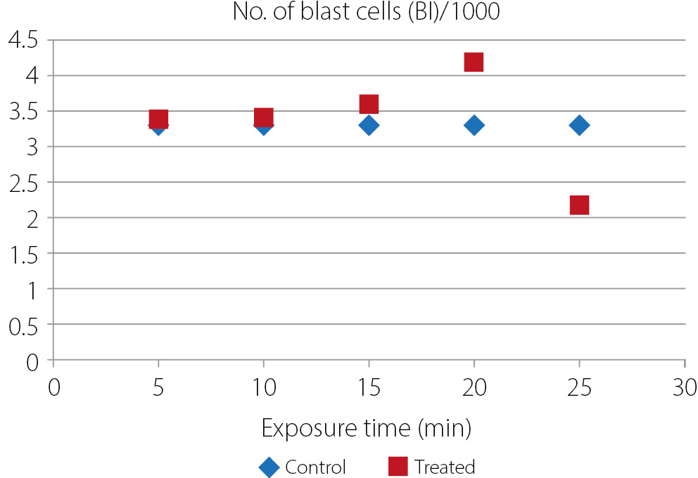
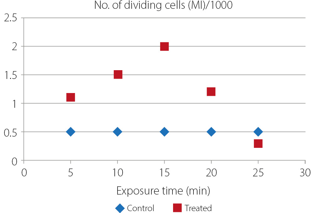
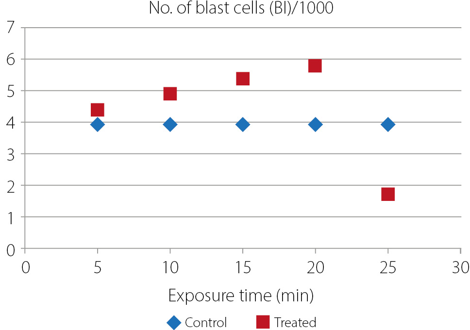
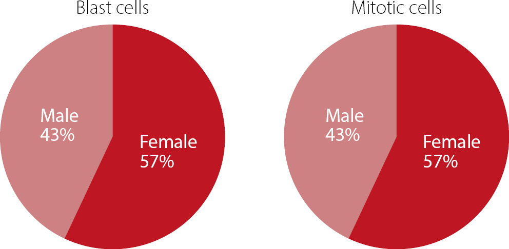
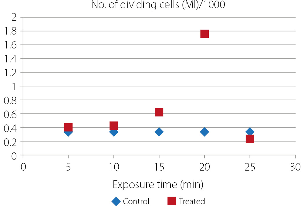
The results demonstrated that short-term exposure of lymphocytes to cold plasma, ranging from 5 min to 20 min, significantly promoted lymphocyte proliferation. This enhancement was evidenced by an increased number of cells primed for division, leading to a higher proportion of actively dividing cells. These observations are supported by the quantitative data presented in the corresponding tables and figures. In contrast, prolonged plasma exposure of 25 min resulted in an inhibitory effect on lymphocyte proliferation. This suppression was characterized by a marked decrease in the number of cells prepared for mitosis, with values significantly lower than those observed in the control group. These findings provide scientific and medical insights, highlighting that plasma can exert beneficial effects in both scenarios, depending on the type of cells being treated. In the case of healthy lymphocytes, short-term plasma exposure appears to enhance cell proliferation, thereby strengthening and supporting the immune response. Conversely, in diseased or abnormal cells, prolonged plasma exposure (≥25 min) may exert inhibitory effects, slowing down or preventing pathological cell proliferation. As illustrated in the figures, the enhanced effect of cold plasma observed in female lymphocytes may be attributed to the interaction between female hormones, cell membrane composition, and a relatively stronger immune response, resulting in increased sensitivity to plasma-generated reactive species. Molecular techniques can be utilized to investigate the effects of cold plasma on target cell genes and related molecular pathways. Such molecular biological approaches have been widely applied across numerous experimental studies in the life sciences in general and in medical research in particular.26, 27, 28, 29

Limitations

The current investigation was constrained by the limited sample size and the exclusive use of in vitro experiments. The molecular mechanisms related to plasma-induced cellular responses were not examined. Additional research with more extensive experimental models is necessary to validate the results.

Conclusions

Accordingly, it can be concluded that brief plasma exposure is beneficial for supporting normal cellular activity, whereas longer exposure durations may be more suitable for the therapeutic targeting of diseased cells. The biological effects of cold plasma are primarily mediated through the generation of reactive oxygen and nitrogen species (ROS and RNS), which interact with cellular membranes and proteins, leading to alterations in cell viability and immune activity.

Data availability

The datasets generated and/or analyzed during the current study are available from the corresponding author on reasonable request.

Consent for publication

Not applicable.

Use of AI and AI-assisted technologies

Not applicable
Tables


Table 1. Mitotic and blastogenesis index of lymphocyte cells at 25 min exposure time

Number of samples

Control

Treated

Male group

mitotic index ±SD

blastogenesis index ±SD

mitotic index ±SD

blastogenesis index ±SD

1

0.4 ±0.12

4.2 ±0.22

0.3 ±0.20

3.3±0.11

2

0.5 ±0.03

2.2 ±0.13

0.4 ±0.10

1.5±0.12

3

0.3 ±0.12

3.5 ±0.12

0.2 ±0.12

2.1±0.11

4

0.2 ±0.08

3.5 ±0.09

0.2 ±0.08

2.2 ±0.22

5

0.4 ±0.09

3.5 ±0.19

0.3 ±0.19

2.1±0.21

6

0.4 ±0.46

3.8 ±0.46

0.3 ±0.46

2.9 ±0.11

7

0.4 ±0.45

3.2 ±0.16

0.2 ±0.25

2.1 ±0.13

8

0.2 ±0.03

2.5 ±0.03

0.1 ±0.13

1.1 ±0.10

9

0.3 ±0.46

2.5 ±0.46

0.2 ±0.21

1.3 ±0.16

10

0.2 ±0.09

4 ±0.99

0.1 ±0.19

3.1 ±0.20

Average

0.33±0.05

3.29

0.23±0.06

2.17 ±0.05

SD – standard deviation.
Table 2. Mitotic and blastogenesis index of lymphocyte cells at 20 min exposure time

Number of samples

Control

Treated

Male group

mitotic index ±SD

blastogenesis index ±SD

mitotic index ±SD

blastogenesis index ±SD

1

0.4 ± 0.12

4.2 ±0.22

1.0 ±0.12

5.2 ±0.12

2

0.5 ± 0.03

2.2 ±0.13

0.8 ±0.03

3.2 ±0.03

3

0.3 ± 0.12

3.5 ±0.12

0.7 ±0.12

4.5 ±0.12

4

0.2 ± 0.08

3.5 ±0.09

1.0 ±0.09

3.5 ±0.09

5

0.4 ± 0.09

3.5 ±0.19

0.9 ±0.09

4.5 ±0.09

6

0.4 ± 0.46

3.8 ±0.46

0.4 ±0.46

4.8 ±0.46

7

0.4 ± 0.45

3.2 ±0.16

0.85 ±0.46

4.2 ±0.46

8

0.2 ± 0.03

2.5 ±0.03

0.9 ±0.03

3.5 ±0.03

9

0.3 ± 0.46

2.5 ±0.46

0.9 ±0.46

3.5 ±0.46

10

0.2 ± 0.09

4 ±0.99

1.1 ±0.09

5 ±0.09

Average

0.33 ±0.05

3.29 ±0.03

1.76 ±0.12

4.19 ±0.12

SD – standard deviation.
Table 3. Mitotic and blastogenesis index of lymphocyte cells at 15 min exposure time

Number of samples

Control

Treated

Male group

mitotic index ±SD

blastogenesis index ±SD

mitotic index ±SD

blastogenesis index ±SD

1

0.4 ±0.12

4.2 ±0.22

0.8 ±0.12

4.5 ±0.22

2

0.5 ±0.03

2.2 ±0.13

0.8 ±0.03

2.5 ±0.13

3

0.3 ±0.12

3.5 ±0.12

0.7 ±0.12

3.8 ±0.12

4

0.2 ±0.08

3.5 ±0.09

0.6 ±0.09

3.9 ±0.09

5

0.4 ±0.09

3.5 ±0.19

0.4 ±0.09

3.6 ±0.19

6

0.4 ±0.46

3.8 ±0.46

0.4 ±0.46

3.9 ±0.46

7

0.4 ±0.45

3.2 ±0.16

0.8 ±0.46

3.7 ±0.16

8

0.2 ±0.03

2.5 ±0.03

0.6 ±0.03

2.9 ±0.03

9

0.3 ±0.46

2.5 ±0.46

0.5 ±0.46

2.9 ±0.46

10

0.2 ±0.09

4 ±0.99

0.6 ±0.09

4.7 ±0.99

Average

0.33±0.05

3.29 ±0.06

0.62 ±0.04

3.6 ±0.03

SD – standard deviation.
Table 4. Mitotic and blastogenesis index of lymphocyte cells at 10 min exposure time

Number of samples

Control

Treated

Male group

mitotic index ±SD

blastogenesis index ±SD

mitotic index ±SD

blastogenesis index ±SD

1

0.4 ±0.12

4.2 ±0.22

0.6 ±0.12

4.3 ±0.32

2

0.5 ±0.03

2.2 ±0.13

0.5 ±0.03

2.3 ±0.23

3

0.3 ±0.12

3.5 ±0.12

0.3 ±0.12

3.6 ±0.22

4

0.2 ±0.08

3.5 ±0.09

0.2 ±0.09

3.6 ± 0.1

5

0.4 ± 0.09

3.5 ±0.19

0.4 ±0.09

3.6 ±0.19

6

0.4 ±0.46

3.8 ±0.46

0.4 ±0.46

3.9 ±0.16

7

0.4 ±0.45

3.2 ±0.16

0.4 ±0.46

3.4 ±0.26

8

0.2 ±0.03

2.5 ±0.03

0.2 ±0.03

2.6 ±0.13

9

0.3 ±0.46

2.5 ±0.46

0.4 ±0.46

2.6 ±0.12

10

0.2 ±0.09

±0.99

0.2 ±0.09

4.5 ±0.99

Average

0.33 ±0.05

3.29

0.4 ±0.06

3.4 ±0.99

SD – standard deviation.
Table 5. Mitotic and blastogenesis index of lymphocyte cells at 5 min exposure time

Number of samples

Control

Treated

Male group

mitotic index ±SD

blastogenesis index ±SD

mitotic index ±SD

blastogenesis index ±SD

1

0.4 ±0.12

4.2 ±0.22

0.5 ±0.11

4.5 ±0.22

2

0.5 ±0.03

2.2 ±0.13

0.5 ±0.01

2.2 ±0.13

3

0.3 ±0.12

3.5 ±0.12

0.5 ±0.11

3.5 ±0.12

4

0.2 ±0.08

3.5 ±0.09

0.4 ±0.19

3.5 ±0.09

5

0.4 ±0.09

3.5 ±0.19

0.6 ±0.09

3.5 ±0.19

6

0.4 ±0.46

3.8 ±0.46

0.4 ±0.26

3.8 ±0.46

7

0.4 ±0.45

3.2 ±0.16

0.4 ±0.16

3.2 ±0.16

8

0.2 ±0.03

2.5 ±0.03

0.3 ±0.13

2.5 ±0.03

9

0.3 ±0.46

2.5 ±0.46

0.5 ±0.11

2.5 ±0.23

10

0.2 ±0.09

4 ±0.99

0.3 ±0.11

4.5 ±0.11

Average

0.33 ±0.05

3.29

0.42 ±0.44

3.37 ±0.23

Figures


Fig. 1. Number of dividing lymphocyte cells for the male group
Fig. 2. Number of blast lymphocyte cells for the male group
Fig. 3. Number of dividing lymphocyte cells for the female group
Fig. 4. Number of blast lymphocyte cells for the female group
Fig. 5. Comparison of the percentage of plasma effects between men and women

References (29)

  1. Muryoush AQ. The effect of cold plasma on pH, creatine, and the concentration of the most trace elements in human’s nails by using X-ray fluorescent method. Iraqi J Sci. 2022;63(5):2057–2062. doi:10.24996/ijs.2022.63.5.21
  2. Guo Y, Zhang X, Xing Y, et al. Atmospheric pressure plasma treatment increases the antioxidant capacity of the serum in vitro. Sci Rep. 2025;15(1):19127. doi:10.1038/s41598-025-03914-8
  3. Deltadahl S, Gilbey J, Van Laer C, et al. Deep generative classification of blood cell morphology. Nat Mach Intell. 2025;7(11):1791–1803. doi:10.1038/s42256-025-01122-7
  4. Merino A, Puigví L, Boldú L, Alférez S, Rodellar J. Optimizing morphology through blood cell image analysis. Int J Lab Hematol. 2018;40(Suppl 1):54–61. doi:10.1111/ijlh.12832
  5. Wu J, Dong L, Xiang J, Di G. Static electric field exposure decreases white blood cell count in peripheral blood through activating hy-pothalamic–pituitary–adrenal axis. Int J Environ Health Res. 2024;34(1):305–315. doi:10.1080/09603123.2022.2148636
  6. Brech A, Kubinyi G, Németh Z, Bakos J, Fiocchi S, Thuróczy G. Genotoxic effects of intermediate frequency magnetic fields on blood leukocytes in vitro. Mutat Res Genet Toxicol Environ Mutagen. 2019;845:403060. doi:10.1016/j.mrgentox.2019.05.016
  7. Heshusius S, Heideveld E, Burger P, et al. Large-scale in vitro production of red blood cells from human peripheral blood mononuclear cells. Blood Adv. 2019;3(21):3337–3350. doi:10.1182/bloodadvances.2019000689
  8. Leon-Ferre RA, Jonas SF, Salgado R, et al. Tumor-infiltrating lymphocytes in triple-negative breast cancer. JAMA. 2024;331(13):1135. doi:10.1001/jama.2024.3056
  9. Nahrendorf M, Ginhoux F, Swirski FK. Immune system influence on physiology. Science. 2025;389(6760):594–599. doi:10.1126/science.adx4380
  10. Zhao H, He Y, Ling X, et al. Profiles of lymphocyte subtypes in HIV‐infected people with different immunologic responses to antiretroviral therapy. J Med Virol. 2024;96(11):e70081. doi:10.1002/jmv.70081
  11. Loureiro A, Pais C, Sampaio P. Relevance of macrophage extracellular traps in C. albicans killing. Front Immunol. 2019;10:2767. doi:10.3389/fimmu.2019.02767
  12. Modern Genetic Analysis. 3rd ed. New York, USA: W.H. Freeman; 2002. ISBN:978-0-7167-4714-7.
  13. Belli M, Tabocchini MA. Ionizing radiation-induced epigenetic modifications and their relevance to radiation protection. Int J Mol Sci. 2020;21(17):5993. doi:10.3390/ijms21175993
  14. Seghatchian J, Amiral J. Spotlight on the current perspectives on applications of human blood cell culture and organoids: Introductory remarks. Transfus Apher Sci. 2020;59(4):102861. doi:10.1016/j.transci.2020.102861
  15. Navakatikian M, Tomashevskaya L. Behavioral and endocrinal effects of microwaves of non-thermal intensity. In: Carpenter DO, Aĭrape-ti︠a︡n SN, eds. Biological Effects of Electric and Magnetic Fields. Vol 1. San Diego, USA: Academic Press; 1994:236–239. ISBN:978-0-12-160261-1, 978-0-12-160262-8.
  16. Gao S, Yang SH, Wang HY, Wang GS, Yin PG. Excellent electromagnetic wave absorbing properties of two-dimensional carbon-based nanocomposite supported by transition metal carbides Fe3C. Carbon. 2020;162:438–444. doi:10.1016/j.carbon.2020.02.031
  17. Santini R, Santini P, Danze JM, Le Ruz P, Seigne M. Investigation on the health of people living near mobile telephone relay stations: I/Incidence according to distance and sex [in French]. Pathol Biol (Paris). 2002;50(6):369–373. doi:10.1016/s0369-8114(02)00311-5
  18. Tamarin RH. Principles of Genetics. 7th ed. Boston, USA: McGraw-Hill; 2004. ISBN:978-0-07-124320-9.
  19. Moraes AP, Vale AAD, Santos RR, et al. Chromosome number overview in Habenaria (Habenariinae, Orchideae, Orchidaceae): a new smallest diploid number and insight on chromosome number evolution in a karyotypically variable genus [published online as ahead of print on November 18, 2025]. Botanical Journal of the Linnean Society. 2025. doi:10.1093/botlinnean/boaf088
  20. Matsuda Y, Uchimura A, Satoh Y, et al. Spectra and characteristics of somatic mutations induced by ionizing radiation in hematopoietic stem cells. Proc Natl Acad Sci U S A. 2023;120(15):e2216550120. doi:10.1073/pnas.2216550120
  21. Claessen MJAG, Yagci N, Fu K, et al. Production and stability of cultured red blood cells depends on the concentration of cholesterol in culture medium. Sci Rep. 2024;14(1):15592. doi:10.1038/s41598-024-66440-z
  22. Chen Z, Liu P, Xia X, Cao C, Ding Z, Li X. Low ambient temperature exposure increases the risk of ischemic stroke by promoting platelet activation. Sci Total Environ. 2024;912:169235. doi:10.1016/j.scitotenv.2023.169235
  23. Mahon CR, Lehman DC, eds. Textbook of Diagnostic Microbiology. 6th ed. St. Louis, USA: Elsevier Saunders; 2019. ISBN:978-0-323-48218-9.
  24. Rosenberg SA. Lymphocytes as a living drug for cancer. Science. 2024;385(6704):25–26. doi:10.1126/science.adp1130
  25. Abdullah MM, AL-Rubaii BAL. Effect of Lactobacillus supernatant on swarming-related gene expression in Proteus mirabilis isolated from urinary tract infections. Ukr J Nephrol Dial. 2024;4(84):39–48. doi:10.31450/ukrjnd.4(84).2024.05
  26. Ibrahim GJ, Laftaah BA. The efficiency of certain amino acids in regulating chABCI gene expression in Proteus mirabilis. Iraqi J Sci. 2024;65(9):4983–4992. doi:10.24996/ijs.2024.65.9.15
  27. Jassim TS, Talib SS, Jaber NR, Sahib DH, Ali RW, Al-Rubaii BA. Impact of hepatitis C virus on IFITM3 gene expression: A comprehensive analysis incorporating serological detection and viral load quantification via qPCR. Polim Med. 2025;55(1):21–30. doi:10.17219/pim/199333
  28. Khudair S, Al-Rasheed IA, Ibrahim TK, Saleh TH, Al-Rubaii BA. Molecular study of CACNA1A, ATP1A2, and SCN1A genes and its association with the migraine disease in Iraq. Ibom Med J. 2026;19(1):132–143. doi:10.61386/imj.v19i1.922
  29. Baqey DAASA, Sultan RS, Hamoode RH, Al-Rubaii BAL. Metabolic markers of insulin resistance in Iraqi type 2 diabetes mellitus patients infected with H. pylori. East Ukr Med J. 2025;13(2):417–423. doi:10.21272/eumj.2025;13(2):417-423